Oak Brook iCAT Offers a Cutting Edge Approach to Dental Implant Diagnosis and Treatment Planning
Oak Brook iCAT is the first dental imaging center of its kind to find a home in the suburbs of Chicago for dental specialists and dentists to diagnose, treatment plan, and follow their dental implant patients.
In today's day and age, diagnosis, imaging, comprehensive medical history, and treatment planning need to be thoroughly evaluated before placement of any dental implants. All of these factors help decrease risk to both the patient and the doctors placing and restoring dental implants.
Imaging is a vital component of the diagnosis, treatment planning, and follow up for any patient undergoing dental implant treatment. The only way to have complete safety and security is with a CT scan prior to placement of dental implants and during follow-up care.
The i-CAT is the state-of-the-art in dental digital imaging technology today. It delivers imaging quality demanded by today's practitioners. Benefits of the i-CAT Cone Beam 3-D Imaging System include:
- Immediate 3-Dimensional images of a patient's critical anatomy
- Complete views of all oral structures allowing dentists & specialists to dramatically enhance patient care
- Open Environment seated position
- Only an 8.5 second scan time
- Processing time in under 1 minute
- Dramatically lower radiation compared to traditional medical CT scans
- Immediate 3-D reconstruction of a patient's mouth, face, and jaw
- Enhances communication between the doctor and the patient
- Allows doctors to share a visual diagnosis with their patients
- Patients better understand their treatment options
- Patients have more confidence going into treatment
- 3-D Mapping Tools allow specialists and technicians to treatment plan more involved situations
Now oral surgeons, periodontists, oral implantologists, and dentists in the suburbs have a place for their patients to get CT Scans in the west suburbs: Oak Brook iCAT.
Oak Brook iCAT Imaging Center is located in Oakbrook Terrace, Illinois and provides patients and referring doctors with a quality CT scan on disk. This is a new standard of care for the periodontists, oral surgeons, oral implantologists, and dentists. The i-CAT allows dental specialists and dentists to obtain a 3-Dimensional view of specific targeted regions for diagnostic and treatment planning purposes.
Traditional Cat scans have been available at hospitals and x-ray centers for many years, but those systems emit a very large dose of radiation. The i-CAT emits a dose less than equivalent of 4 days of standard background radiation. The scan time for the i-CAT is 8.5 seconds, which is also much shorter than traditional CT scans. An astounding amount of information is given in one scan including a panoramic view, a cephalometric view, 3D rendering, TMJ evaluation, and airway evaluation.
With an i-CAT CT scan, skeletal, hard tissue (teeth), soft tissue and other anatomical features can be studied in great detail after just an 8.5 second scan with less radiation than a complete full mouth dental x-ray series.
Oak Brook iCAT scan center is run by Dr. Umar Haque, an oral implantologist and dental radiologist at Oak Brook Smiles. Dr. Haque is a Fellow in the International Congress of Oral Implantologists in both the prosthetic and surgical divisions and a Fellow in the American Dental Implant Association. Dr. Haque added, "The i-CAT’s high resolution, volumetric images provide complete 3-D views of critical anatomy for more thorough analysis of bone structure and tooth orientation to optimize implant treatment and placement, and selection of the most suitable implant type, size, location, and angulations prior to surgery." Anyone considering dental implant therapy should definitely ask their dentist or specialist if a CT scan will be used for diagnosis, treatment planning, and follow-up care.
Oak Brook i-CAT is located at 1S132 Summit Avenue, Suite 200 in Oakbrook Terrace, Illinois. They can be reached at 630-627-2520 or 630-627-7420, and the scan center is also online at www.oakbrookicat.com
###
Siraj Haque
630-627-2520
www.oakbrookicat.com
630-627-7420 Office
630-396-3863 Fax
630-329-6900 Mobile
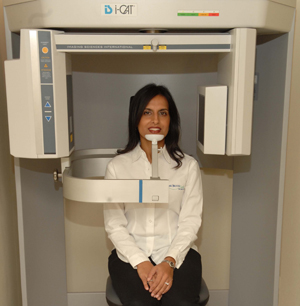
View of the i-CAT at Oak Brook iCAT
Siraj Haque, the Office Manager at Oak Brook iCAT, tests out the CT Scanner.
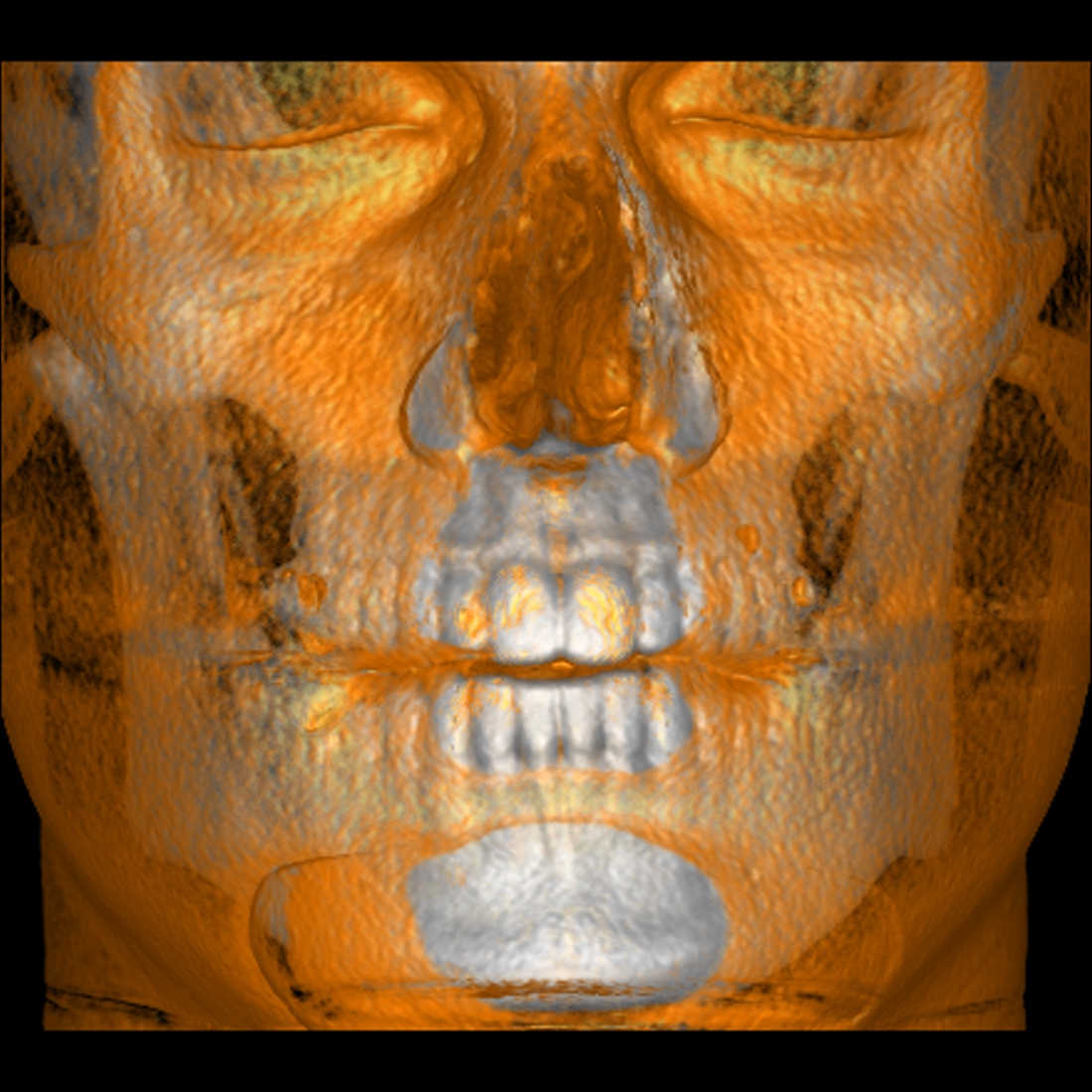
Soft Tissue 3D Rendering of a Patient at Oak Brook iCAT
The ability of the i-CAT to not only see hard tissues such as teeth and bone, but also soft tissues in relationship to them.
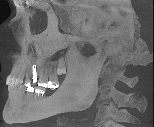
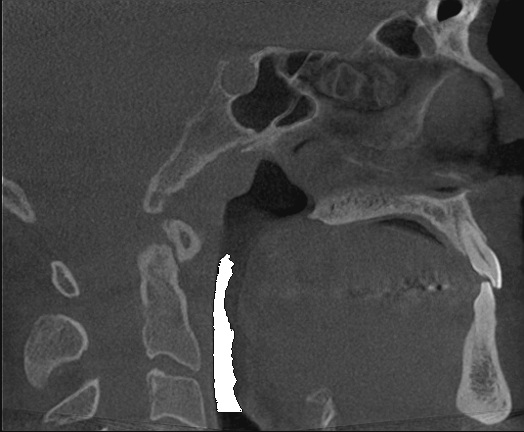
A Cephalometric View of an Implant Patient
Many cross sections are taken during a scan, and a Cephalometric view is also obtained during a full scan.
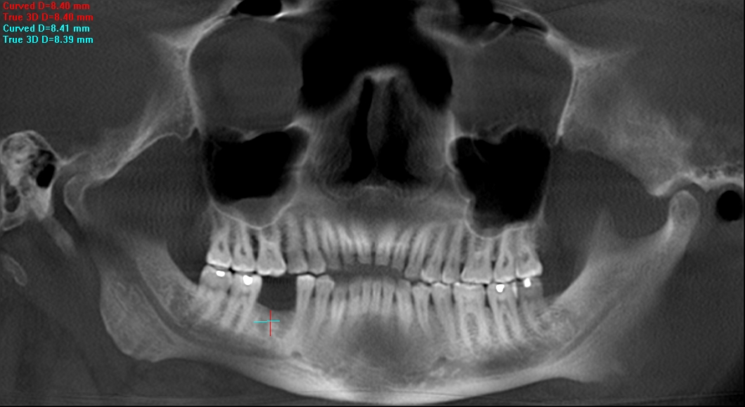
A Panoramic View also is obtained during a scan
The i-CAT also renders a beautiful panoramic view of a patient, showing the jaws, teeth, TMJ, and maxillary sinuses. Note the swelling in the right sinus (left side of screen) and exact 1:1 measurements for initial implant planning.

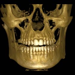
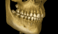
3D Rendering of Bone and Teeth
From a scan, one can also render just the hard tissues.
Airway Evaluation from an i-CAT CT Scan
One can also get a nice evaluation of the airway, as is the case for this patient with obstructive sleep apnea.
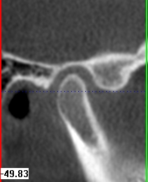
Wisdom Teeth in 3D
Rendering of wisdom teeth is another added bonus with an i-CAT Scan.
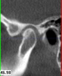
Views of the TMJ: An example of a healthy joint
Cross sectional views of the TMJ once required a few separate scans. Now it is all available in one to two scans. Here is an example of a healthy TMJ.
Views of the TMJ: An example of a deteriorating joint
Cross sectional views of the TMJ once required a few separate scans. Now it is all available in one to two scans. Here is an example of a deteriorating TMJ. This is the same patient, but the opposing symptomatic, painful side.