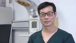
International Standards and Guidelines for Neonatal Lung Ultrasound by a Group of International Experts from Oasis Publishers

New York, NY, March 25, 2019 --(PR.com)-- An international standards and Guidelines for Neonatal Lung Ultrasound titled, “Protocol and Guidelines for Point-of-Care Lung Ultrasound in Diagnosing Neonatal Pulmonary Diseases Based on International Expert Consensus” was published recently in the Journal of Visualized Experiments (doi:10.3791/58990) by a group of international experts.
It was said that in the department of neonatology and NICU of Beijing Chaoyang District Maternal and Child healthcare Hospital since March 2017, chest X-rays have been replaced comprehensively by lung ultrasounds and are routinely used for the diagnosis and differential diagnosis of neonatal lung diseases. Thus, the main objective of the protocol is to popularize the application of point-of-care lung ultrasounds (POC-LUS) in neonatal intensive care units (NICUs).
According to the U.S. National Library of Medicine, an ultrasound is a type of imaging, which uses high-frequency sound waves to look at organs and structures inside the body. Since the ultrasound is the safest imaging tool, it “obviates the use of ionizing diagnostic procedures.” Due to its convenience, it has received increasing attention from neonatal physicians. However, the need to follow clear reference standards and guideline limits has been felt amongst the physicians and researcher communities.
Dr. Liujing and an international expert team observed that despite the chest x-ray being used as the main imaging tool in the diagnosis of lung diseases, yet the lung ultrasound was considered a “forbidden zone” in the detection of lung diseases. This was because ultrasonic waves are reflected when encountering air. However, utilizing artefacts formed by different pathological changes in adults, children and new-born infants challenged this understanding.
Nonetheless, the use of point-of-care lung ultrasound remains limited and to promote proper utilization of POC-LUS in the neonatal field, an international expert panel to review the latest publications on neonatal LUS was set up. The panel summarized these expert opinions and developed the present LUS protocols and guidelines for its use. Dr. Liu suggests that as a real imaging technique, LUS is user-friendly, easy to learn and easy to replicate with appropriate training.
Dr. Liujing and international expert team followed a certain protocol and the main purpose of the protocol and the guideline is to instruct users on how to use LUS to diagnose and differentiate common neonatal lung diseases. These include respiratory distress syndrome (RDS), transient tachypnoea of the new-born (TTN), pneumonia, meconium aspiration syndrome (MAS), pulmonary haemorrhage, pulmonary atelectasis and pneumothorax, etc. The normal neonatal characteristics and the LUS diagnostic criteria for different lung diseases have been described in detail in the research paper.
The team of researchers observed that POC-LUS is a feasible and convenient diagnostic method that can be performed in the neonatal intensive care unit at the bedside. Furthermore, it is a very sensitive and reliable method in the diagnosis of all types of neonatal lung diseases. Liu and the team concluded that it has multiple advantages over the chest x-ray (CXR) and/or chest computerized tomography (chest CT) scan such as accuracy, low-cost, reliability, simplicity and no risk of adverse effects due to radiation.
The guidelines have encouraged the use of LUS in the neonatal intensive care unit. However, their findings suggest that while studying the imaging modality, some issues should be considered, the details of which can be found in the research paper. According to Dr. Liujing and the team of researchers, perpendicular scanning is the most important and most commonly used scanning method. This type of scanning can also be used to examine the integrity of the diaphragm and the presence of pleural effusions.
However, the team advises that in clinical practice, LUS should not be limited to a fixed scanning sequence. Occasionally, the use of extended view function (XTD-View) can be brought about in practice. In addition to the above, Dr. Liujing and the team of researchers mention caveats. The LUS has limitations such as its being highly operator dependent and therefore the need for sufficient experience to fully understand the principles of LUS before conducting examinations. Another limitation is that some mild cases may be missed if the scan is not performed carefully. Some other limitations have been mentioned as well.
According to Liu, current literature offers well-designed, systematic and in-depth research in the area of LUS. Research findings have been validated and confirmed in clinical practice. Furthermore, Liu mentions that the protocol and guidelines followed during their research have been developed after a thorough evidence-based review of the available data by a panel of international experts in this field.
In-Depth research by Dr. Liujing and international expert team display their understanding and analytical skills in this field. Their hard work must be applauded as their research work sets a positive precedent in the field of clinical research. It can be a game changer in the medical world and can change the way point-of-care lung ultrasound diagnosis various neonatal lung diseases.
Contact
Oasis Publishers
Jing Liu
+1-646-751-8810
www.oasispub.org
Jing Liu
+1-646-751-8810
www.oasispub.org
Categories
